Summary
With the right AI radiology solution, healthcare providers spend less time on repetitive image review operations. Further, software automatically detects patterns, improves diagnostic confidence and streamlines reporting workflow. In this blog, I will discuss how AI-powered radiology software helps clinics take timely and more confident decisions. Also, I will discuss how AI routine image interpretation handles workload in a frictionless way, especially in high-volume setups.
Introduction
Clinics can clearly visualize that the radiology department is facing huge challenges in 2026. Imaging volumes are consistently increasing, reporting timelines are becoming tight, and there is zero compromise in experience in diagnostic accuracy. In this context, AI-powered radiology software makes the image analysis faster and more efficient. Traditionally, radiologists are highly dependent on manual images, which increases the risks of fatigue, time pressure, and repetitive scan errors. If you view it in a bigger perspective, an AI-powered radiology information system is not just a tech upgrade; it has become a long-term clinical and operational strategy. Keep reading!!!
Core Technologies Behind AI Radiology
Let’s see:
Deep Learning Models Behind Intelligent Imaging
Most AI Radiology technologies have been developed based on Deep Learning Models. They have a very valid construction such that they are trained on huge worksheets to establish and discover extremely complicated patterns that are invisible to human eyes. They evaluate medical images and then signal the hidden and very subtle clinical indicators in them.
CNNs for High-Precision Image Segmentation
Convolutional Neural Networks are those for deep segmentation of images. Moreover, it will define any abnormality, such as tumors, fractures, or even those minute anomalies found in the view. In addition, this model can easily obtain above 0.98 AUC scores for diagnostics, which speaks volumes about reliability as well as consistency in diagnosis.
Seamless Integration with PACS and RIS Systems
The latest AI radiology solutions integrate with the existing PACS or RIS systems so smoothly that the workflow becomes absolutely complication-free. Whether a cloud-based deployment or an on-premise setup is concerned, the AI tools will always run in the background and provide the radiologists with faster access and analysis support.
NLP-Driven Reporting & Time Savings
Natural Language Processing (NLP) makes the imaging workflow more efficient, as it automatically generates customized radiology reports from dictated findings. Furthermore, this AI-assisted reporting minimizes repetitive work and saves valuable time for radiologists. Tools like healthray, save 60+ minutes of reporting time in just one shift. Further, this shifts the focus of radiologists to diagnosis and patient care while significantly reducing the workflow errors or delays.
Multimodal AI for Predictive Insights
Advanced multimodal AI combines imaging data with EHR information. Further, radiology information software enables predictive analytics such as radiomics-based risk scoring. This approach is not just limited to current findings but it also strengthens future risk assessment or clinical decision-making.
Addressing Key Challenges in AI Radiology Adoption
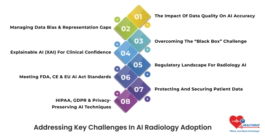
AI radiology delivers significant value but there are some important challenges that come along with real-world deployment and that need proactive consideration. Let’s find out:
The Impact of Data Quality on AI Accuracy
AI model accuracy largely depends on training data quality. Furthermore, if the dataset is limited or homogeneous, then predictions become clinically unreliable.
Managing Data Bias & Representation Gaps
Biased datasets make the AI outputs unreliable, especially when diverse patient populations are involved. Furthermore, real-world, multiethnic, and geographically diverse data increases the importance of training in order to ensure inclusivity and consistency.
Overcoming the “Black Box” Challenge
Majority of AI radiology systems function as a “black box,” where prediction logic is not transparent. Further, this lack of visibility creates trust issues for radiologists.
Explainable AI (XAI) for Clinical Confidence
Explainable AI (XAI) helps radiologists visualize which features and image regions influence the final prediction. Further, it improves the decision-making confidence and makes the AI adoption smooth.
Regulatory Landscape for Radiology AI
Radiology AI solutions Software comes under the category of Medical Device (SaMD), where regulatory compliance is non-negotiable.
Meeting FDA, CE & EU AI Act Standards
Frameworks like the FDA, CE marking, and the upcoming EU AI Act make the safety, accountability, and data traceability protocols requisite before clinical implementation.
Protecting and Securing Patient Data
As we know, healthcare data are highly sensitive; therefore, privacy compliance is essential for AI radiology platforms.
HIPAA, GDPR & Privacy-Preserving AI Techniques
To follow HIPAA or GDPR regulations, AI vendors adopt federated learning and privacy-preserving techniques, where models are trained on decentralized data without transmitting the patient data.
Core Purpose & Functionality of AI in Radiology
Let’s check out:
Rising Imaging Volumes & Manual Review Pressure
In today’s radiology department, imaging volumes are rapidly increasing. Further, radiologists have to daily review hundreds of images manually, which is time-consuming and increases the risks of exhaustion!.
Risk of Missed Findings Due to Workload
High workloads increase the likelihood of overlooking subtle abnormalities, such as small fractures or early-stage lesions. Further, these delays negatively impact diagnosis and patient outcomes.
AI-Driven Automated Detection & Interpretation
The primary role of AI in radiology is automated detection or interpretation. Additionally, researchers train models on vast datasets of labeled medical images. This helps clinics to quickly and accurately diagnose conditions such as pulmonary nodules, intracranial hemorrhages, breast lesions, bone fractures, and vascular blockages.
Faster Identification of Critical Abnormalities
AI algorithms prioritize critical findings that highlight urgent cases beforehand. Further, this minimizes the reporting turnaround time and creates possibilities of timely clinical intervention.
Advanced Image Reconstruction & Quantitative Analysis
Beyond detection, AI-powered radiology software supports AI image reconstruction and quantitative analysis. Further, it provides precise measurements and objective insights to radiologists. Thereby, improving the diagnostic confidence.
Automated Reporting to Reduce Repetitive Work
AI-assisted report generation significantly reduces repetitive manual documentation. Further, structured and consistent reports reduce the administrative burden of radiologists.
Enabling Radiologists to Focus on Clinical Decisions
As the pressure of manual reviews and documentation workloads minimizes, radiologists can shift their focus to high-value clinical decision-making and patient care without compromising quality,speed and accuracy.
 PRO TIP
How AI Works in Image Analysis
Ever wondered how AI radiology software works? What happens as the image gets loaded into the system? And how does AI fetch the meaningful insight in just a few seconds? Let’s check out:
Step 1: Data Acquisition: Where Does the Image Come From?
The AI journey begins from the imaging machine. Further, digital images from CT, MRI, PET, and X-ray systems directly flow into the AI pipeline. Now the question is, “Do all of the images have the same format and quality?” Not really; that’s the reason the next step is highly critical.
Step 2: Preprocessing: How does AI Make the Images Clean and Standard?
In the processing stage, AI software enhances the image quality, removes unwanted voices, and standardizes the format. Further, these steps ensure the algorithm works on clear and consistent images. Advanced AI tools can enhance images by adjusting contrast, sharpening details, and removing noise, ensuring radiologists work with the clearest possible visuals for accurate diagnosis. After all, if input is unclear, how will output get accurate?
Step 3: Segmentation & Detection: How Does the System Identify Anomalies?
In this stage, AI algorithms segment anatomical structures and detect abnormal patterns. Further, AI efficiently scans each critical area of the tumor, fracture, and vascular blockage. Do you think human eyes can work with the same speed or consistency? It is practically impossible!!!
Step 4: Classification & Reporting: How Findings Become Actionable
In the last stage, systems classify the detected findings and generate preliminary reports to keep the radiologist validation at hand. Moreover, this makes the reporting faster, and radiologists get structured insights, preventing repetitive manual work.
Faster Processing, Zero Fatigue
Through this structured pipeline, AI processes the massive imaging volumes with higher speed, beyond the human capacity, which results in faster diagnosis, better efficiency, and improved patient outcomes.
 NOTE
NOTE
Emerging Trends in AI Radiology
AI radiology is not just limited to abnormality detection. The real question is how software makes the diagnosis predictive, personalized, and/or proactive. Let’s find out:
Multimodal Data Fusion
- When Imaging Meets Complete Patient Data
Future AI models will combine imaging data with EHRs, lab results, and genomics. Further, this opens possibilities of integrated diagnostics that will highlight both present conditions and future risks. That means the doctor will get a complete picture of both the problem and the solution.
- Why are personalized insights important?
Every patient is different; when AI understands the entire patient medical history, then treatment becomes more accurate and personalized, thereby eliminating improvisation.
Automated Image Quality Control
- Catching Scan Errors Before They Cause Delays
Scans are sometimes repeated because the images are blurry or the patient’s positioning was incorrect. Further, AI detects patient motion, wrong positioning, and underexposure issues at the time of scans, before finalizing the images.
- Reducing Repeat Scans & Workflow Disruptions
This automation reduces the repeat scans and reporting delays and radiology workflows become less stressful.
Cloud-Native & Edge Diagnostics
- Collaborative Reading Across Hospital Networks
Cloud-native platforms provide options for radiologists to review the scans of multiple locations. Thus, it enables shared expertise.
Step towards digital era with our healthcare solution
Revamp your hospital facilities and embrace change for better healthcare management. Ease in managing and organizing large medical datasets leads to effective analysis. Seize the opportunity now!
- Edge AI for Rural & Mobile Screening Units
When connectivity and specialist availability are scarce, Edge AI offers immediate access to remote locations and mobile diagnostic units.
Real-Time 3D Visualization & AR Integration
- From Flat Images to Immersive 3D Models
AI-powered 3D visualization converts radiological images to complete anatomical models.
- AR/VR for Surgical Planning & Simulation
AR/VR technologies allow surgeons to simulate complex procedures that minimize intraoperative risks.
Conclusion
AI’s biggest strength is not about replacing radiologists. When AI intelligence and human expertise cooperate, its true worth is revealed. AI algorithms amplify clinical intelligence to help radiologists make confident, timely, and intelligent decisions. Its impact will not just be limited to hospitals; instead, it will expand to global healthcare equity.